“Dopamine Rush” – The Story of Exchange
Dopamine is the main neurotransmitter involved in the pathway that regulates reward-motivated behavior [1]. The rewarding feeling that we experience when spending quality time with our loved ones, or acquiring a perfectly resolved NMR spectrum, comes from the increased dopamine level. This in turn motivates us to repeat the actions in exchange for more rewards. Let’s take a closer look at the molecular level of this small but powerful molecule.
Dopamine, derived from the amino acid L-tyrosine, consists of a 1,3,4-trisubstituted benzene ring with the substituents being two hydroxyl and one ethylamine groups. The 1D1H spectrum of dopamine dissolved in DMSO-d6 shows the signals for the ethyl’s CH2 groups in the 2-3 ppm region, and the protons on the benzene ring in the 6-7 ppm region (Figure 1A). More noticeably, the signals for the protons on the -OH and -NH3+ groups of dopamine can be observed as broad singlets. This broader lineshape is due to the chemical exchange of these protons with one another, and with the residual water in the DMSO-d6 solvent (the singlet at 3.33 ppm).
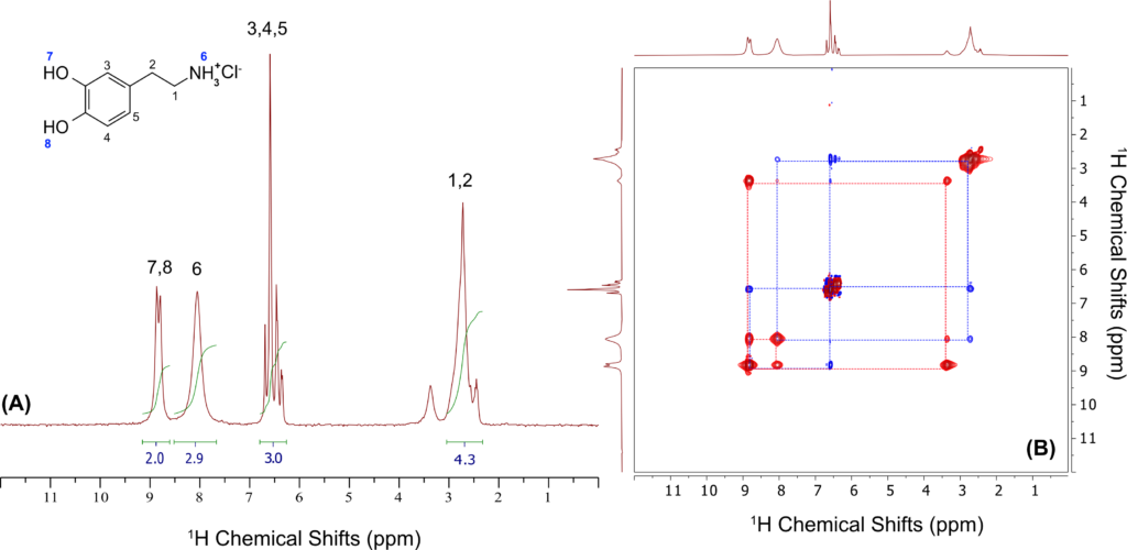
Figure 1. (A) 1D1H NMR spectrum of Dopamine in DMSO-d6; (B) 2D 1H-1H ROESY NMR spectrum of Dopamine acquired using 300 ms mixing time.
Chemical exchange among labile protons can be easily visualized with NMR using either Rotating Frame Overhauser Effect Spectroscopy (ROESY) or Chemical Exchange Saturation Transfer (CEST). A 2D-ROESY NMR spectrum of dopamine is shown in Figure 1B. The ROESY spectrum can reveal correlations due to cross-relaxation including Nuclear Overhauser Effect (NOE), chemical, or conformational exchange [2]. In the ROESY spectrum of dopamine, the cross-diagonal peaks that are in the same phase as the diagonal signals (red) arise from the chemical exchange between the labile -OH and -NH protons and the protons from the water in DMSO. The correlations that are in the opposite phase of the diagonal signals (blue) are due to NOE.
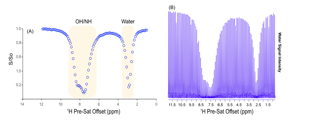
Figure 2. (A) A CEST spectrum corresponds to the response of the water signal due to 1H pre-Saturation offset. (B) A series of 1H NMR spectra showing the response of the water resonance as we sweep through the Pre-SAT offset.
Another method for studying chemical exchange is Chemical Exchange Saturation Transfer (CEST). This technique monitors the response of labile 1H resonances as the pre-saturation frequency is systematically swept across the entire 1H chemical shift range [3]. Figure 2 shows a series of 1H NMR spectra that illustrate this behavior. The response of the water protons signals to variations in the 1H pre-saturation offset enables the tracing of exchangeable 1H sites, producing a silhouette that reflects the labile proton environments within the molecule. Importantly, this approach requires only a single exchangeable site to be resolved and can be used to identify additional exchangeable sites even in the presence of overlapping resonances.
When you exchange chocolate and cards this Valentine’s Day and are experiencing that “dopamine rush”, know that at the molecular level, the labile protons on dopamine are also busy exchanging between different sites. Happy Valentine’s Day!
To read the complete App Note please Click Here
References
[1] K. C. Berridge, “The debate over dopamine’s role in reward: the case for incentive salience,” Psychopharmacology (Berl)., vol. 191, no. 3, pp. 391–431, Mar. 2007, doi: 10.1007/s00213-006-0578-x.
[2] “University of Ottawa NMR Facility Blog: What are Those Positive Peaks in My NOESY Spectrum?” Accessed: Feb. 09, 2026. [Online]. Available: https://u-of-o- nmr-facility.blogspot.com/2008/07/what-are-those-positive-peaks-in-my.html
[3] “University of Ottawa NMR Facility Blog: CEST.” Accessed: Feb. 09, 2026. [Online]. Available: https://u-of-o-nmr-facility.blogspot.com/search/label/CEST