Analytical NMR

NMR is an excellent tool for quantification and identification. The peaks in the spectrum will appear in predictable locations in the spectrum and their intensities are proportional to the number of nuclei in the sample.

Analytical NMR

NMR is an excellent tool for quantification and identification. The peaks in the spectrum will appear in predictable locations in the spectrum and their intensities are proportional to the number of nuclei in the sample.

Identification, Quantification (qNMR) and Mixture Analysis

NMR is best known for its use in structure determination. The chemical structure of a small molecule dictates a rational spectrum. This is because the electronic structure, the bedrock of chemistry, is what determines the chemical shift, while the number of nuclei determine the coupling and relative intensities.

These same properties result in NMR being excellent for quantification and identification. The peaks in the spectrum will appear in predictable locations in the spectrum and their intensities will be proportional to the number of nuclei in the sample. In other words, the peaks are a chemical signature and the sample concentration is proportional to the intensity of the NMR signal detected. Sample concentrations and purities can be easily measured from known peaks once the proportionality constant is calibrated using a reference of known concentration. Using these peak positions and relative intensities, the compounds, mixtures and impurities in a sample can be identified or quantified.

Identification

NMR spectra can be used to identify compounds. Depending on the degree of specificity required, this can be a simple peaks comparison or can incorporate other tests related to peak splitting and integral intensities. Here we show a method for identification of ascorbic acid, which is a common vitamin sold on its own as well as in many dietary supplements.

The United States Pharmacopeia method for identification of ascorbic acid requires both FT-IR and a reducing sugar chemical test because neither has the specificity on its own. Here a single NMR spectrum is acquired and analysed with three different tests: peaks, multiplet, and nuclides count comparisons to quickly identify ascorbic acid. A similar set of tests is used for sodium ascorbate.

Peaks Test

The peak positions in the spectrum are compared to a peaks list determined from a standard material. If the peaks match, then the sample passes the peaks comparison test. The peak positions are shown above the spectrum and range from 3.64 to 4.99 for this sample.

Figure 1: Peak matching of the ID test.

Multiplet test

The multiplet position and multiplicity of the peaks in the spectrum are compared to those determined from a standard material. If the two match, then the sample passes the peaks comparison test. The peak positions and multiplicity are shown in the boxes on the spectrum. Peak A is a doublet (d) at 4.96 ppm and Peak B is a multiplet (m) at 3.85 ppm.

Figure 2: Multiplet matching of the ID test.

The sample must past all three tests to be positively identified as ascorbic acid. This identification test was validated using the following tests for specificity and reproducibility.

TestCategoryResult
Positive identificationSpecificity12 samples resulted in positive IDs
Negative controlSpecificity4 analytes not identified as ascorbic acid
RepeatabilityPrecision6 standards measured in one day
Intermediate PrecisionPrecision3 standards measured over three days
Reproducibility (Ruggedness)Precision6 standards measured in two labs

These results meet the UPS specifications for an identification method.

Quantification – Internal Standard

Quantification using any analytical method is no more than calibrating an instrumental response with a known reference, and then calculating the concentration of an unknown sample from the measured instrument response. One method of calibrating the NMR spectrometer is with an internal standard. Both the sample and the reference are weighed out and co-dissolved into a single solution. The integral of the peak associated with the reference sample is used to calibrate the instrument response. One or more peaks associated with the sample of interest are then used to determine the sample concentration or purity. The purity can be calculated with the following equation.

qNMREquation1

Where:

  • M =Molecular weight
  • I = Integral
  • N = Normalisation factor (the number of nuclei represented by the peak)
  • m = Mass
  • P = Purity
  • Sample = designates the sample of interest
  • Ref = Reference Standard

Methylsulfonyl methane

When quantitative methods are validated there are standard requirements for accuracy, precision, range, and linearity over that range. Table 1 below shows general requirements for a Category I NMR method when measuring a drug substance (there are other specifications for finished products and impurities).

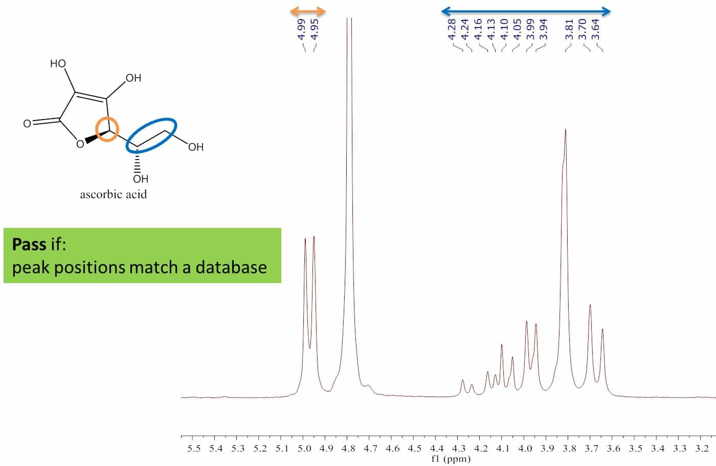
These requirements were tested by measuring the purity of one reference standard, methylsulfonylmethane (MSM), with another, maleic acid. Maleic acid is a common reference standard so this was used as the reference to measure the known purity of MSM (99.5%). A spectrum of the mixture in D2O is shown in Figure 3.

Figure 3: Spectrum of MSM and maleic acid in D2O.

Maleic acid and MSM were co-dissolved in D2O at a concentration of approximately 60 mg/mL. The maleic acid peak is a singlet, has two protons, and has a chemical shift of about 6.3 ppm. The methyl peak of MSM is a singlet representing six protons and located at 3 ppm. The purity was determined from these peaks to assess the accuracy and precision of this method.

Figure 4: Purity for six independently prepared maleic acid and MSM solutions.

Figure 4 shows the purity measured by NMR for six independently prepared maleic acid and MSM solutions. The mean purity and standard deviation measured for MSM was 99.5 ±0.6%. This shows an accuracy within 2% and repeatability (precision) of less than 1%.

Figure 5: The measured MSM purity for 30, 60 and 75 mg/mL (50-120 % of 60 mg/mL).

This experiment was repeated for 30 and 75 mg/mL (50-120% of 60 mg/mL). Over this range the accuracy and precision were 0.9, 0.6, and 0.6%, which is shown in Figure 5.

Figure 6: Linearity check of signal versus concentration.

The linearity test between 15 mg/mL and 75 mg/mL is shown in Figure 4. A correlation coefficient (R2) of 0.9999 was determined, confirming highly linear behaviour between peak integral and concentration.

The results of these experiments are shown in Table 1. This method meets the accuracy, repeatability, range, and linearity requirements for a typical measurement of a drug substance. The uncertainty of these results is for the preparation and measurement of the samples. The error budget for weighing the samples is about 0.3 % or half of the total uncertainty.

 USP SpecificationMeasured
Accuracy98-102 %100 %
Repeatability1 %0.6 %
Range80-120 %50-125 %
Linearity0.9950.999

USP <761> Nuclear Magnetic Resonance / Physical Tests

Table 1: USP requirements for accuracy, repeatability, range and linearity compared to performance of Spinsolve benchtop NMR.

Mixtures Analysis

Common mixtures include solvent systems or block copolymers. NMR is an alternative to common techniques such as chromatography or titration, which can be time intensive and complicated techniques.

Figure 7: Solvent mixture analysis of a Tequila sample.

Alcohol and water is a common solvent mixture. It contains one well resolved peak at about 1.2 ppm and two overlapping peaks between 2.7 and 6.0 ppm. The peak at 1 ppm is from the methyl protons (red box) of the ethanol and the other peaks are from the water protons and the alcohol and methylene protons from ethanol (yellow boxes). The integrals from the peaks can be represented by the following equations:

I1 = 3 EtOH; I2 = 2 H2O + 3 EtOH,

which can be transformed to calculate concentrations of each component using the following equations:

EtOH = I1/3; H2O = (I2 – 3 EtOH)/2.

The volume fraction or percent ethanol is calculated by converting from relative molar fractions to relative volumes using molecular weight and density.

This procedure and calculation was automated and then tested on tequila. After running the protocol we found that it contained 36% alcohol compared to 38% on the label.

Poloxamer

Figure 8: Spectrum of a oxyethylene-oxypropylene block copolymer.

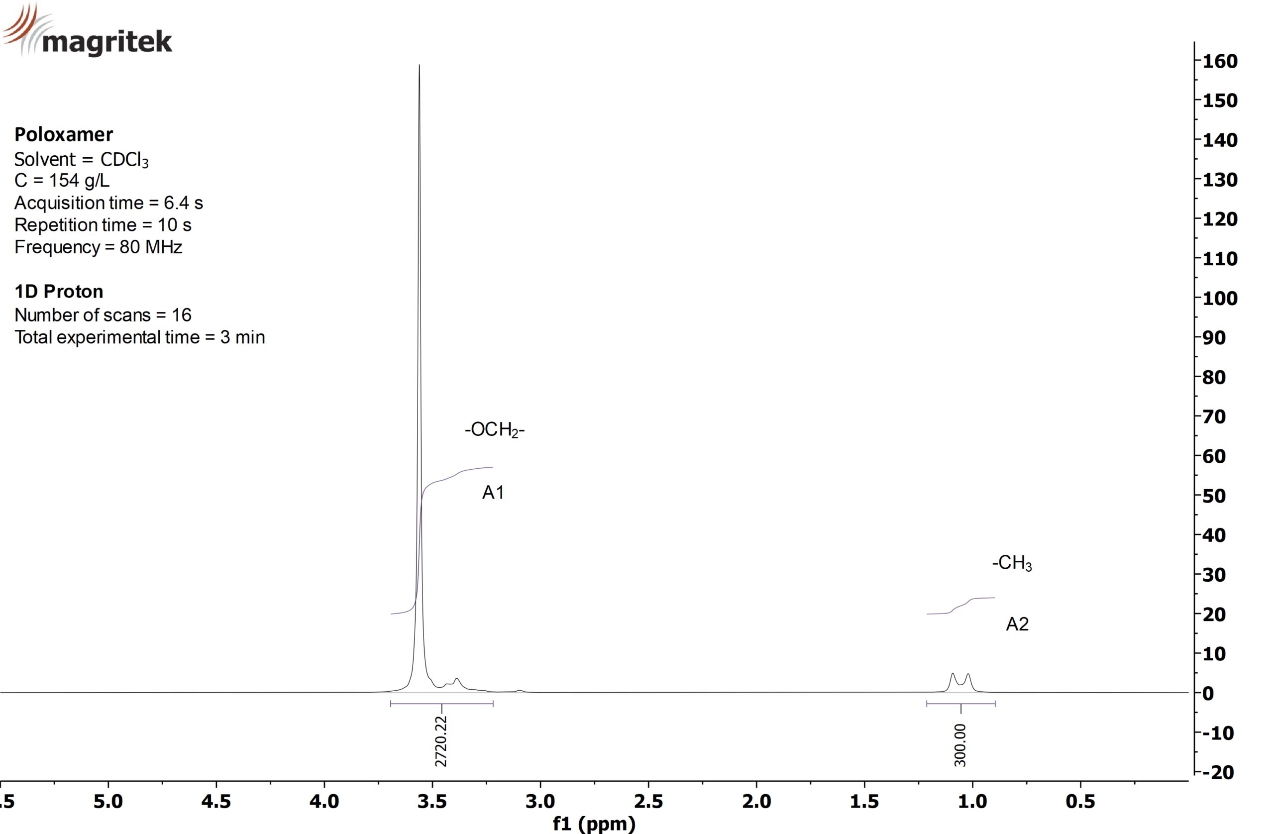
Poloxamers are oxyethylene-oxypropylene block copolymers commonly used as non-ionic surfactants. The ratio of oxyethylene to oxypropylene is a tuneable chemical property that is commonly determined by NMR spectroscopy. The peak at 1 ppm is from the propylene methyl and the peak at 3.5 ppm is from the polymer backbone. The weight percent oxyethylene is calculated using the following equations:

Poloxamer Formel -

The “7” in F127 means that the sample has approximately 70% oxyethylene and the NMR determined it to be 74%.

Examples of 31P NMR

Protons are not the only nuclei that can be used for analytical measurements. Here phosphorous containing acids are used as examples of identification and quantifications using 31P as the observed nucleus.

Identification

In methamphetamine production, red phosphorous is mixed with the precursor, ephedrine or pseudoephedrine. The phosphorus is converted to H3PO2 in the first stage of the reaction and then to H3PO3, and finally converting to H3PO4 at the end of the reaction. The amount of each acid provides an idea of what stage the synthesis was at and the potential yield.

Figure 9: 31P spectra of phosphoric, phosphorous and hypophosphorous acids (left) and mixture (right).

The chemical shift is used to identify the acid.

Quantification

These strong acids are not stable conditions for internal standards therefore an external standard is used. The external standard method involves measuring a sample of known concentration and purity. The sample does not need to be the same as the sample of interest.

Figure 10: Linearity of 31P signal with concentration.

Here you can see the linearity of the NMR integral with hypophosphorous concentration is >0.99 which means you can reliably estimate the concentration across this range.

The 30% hypophosphorous acid sample was used as an external standard for the samples listed in the table below.

SampleTheoretical Concentration (w/w%)Calculated Concentration (w/w%)
Hypophosphorous Acid 15050.76
Hypophosphorous Acid 24040.93
Hypophosphorous Acid 33030.00
Hypophosphorous Acid 42019.67
Hypophosphorous Acid 5109.40
Hypophosphorous Acid Casework Sample 1about 50*52.33
Hypophosphorous Acid Casework Sample 2about 30*18.05

*Case work samples were estimated to be about 30% and 50% hypophosphorous acid using IR spectroscopy.

Table 2: Measurement of hypophosphorous acid concentration using external standard.

The calculated concentration agrees well with the theoretical concentration for the hypophosphorous acid samples.

Internal Standard

Quantitative NMR with internal standard on a Spinsolve benchtop NMR spectrometer.

The knowledge of the assay of a substance is crucial information and of interest in many applications. For example  in pharmaceutical or chemical laboratories, in the production of reference materials, or to follow the conversion of reagents in reactors. In the past decades quantitative nuclear magnetic resonance spectroscopy (qNMR) has been established as a standard method for assay determination, as it takes advantage of the fact that NMR provides a linear signal response. This results in the NMR signal intensity being proportional to the number of nuclei, which allows for an easy one-point calibration against a known standard. The type of qNMR method, which we present in this blog post, utilizes an internal standard as a reference. The other method, using an external standard, will be presented in an upcoming blog post.

For optimum performance of the internal standard qNMR method it is required that the reference material has a known assay, the integrated signals are not overlapping, and that analyte and standard are sufficiently soluble in the desired solvent. One example of this is the determination of the assay of diclofop-methyl with dimethyl sulfone as internal standard. Diclofop-methyl is a post-emergence herbicide used, for example, for wild oats and wild millets. As it can be absorbed by the soil it is important to have a reference material with known assay with which one can determine the contamination of soil sample

blog post

Figure 1:  Structures and molecular weights of diclofop-methyl (left) and dimethyl sulfone (right).

The sample was provided to us by HPC Standards GmbH, a manufacturer and distributor of high-purity analytical standards for residue analysis. For the analysis, a sample of 34.9 mg diclofop-methyl, 4.9 mg dimethyl sulfone and 500 µL methanol-d4 was prepared, homogenized, and transferred into a standard 5 mm NMR tube.

Figure 2 shows the acquired proton spectrum of this sample including the integrals of dimethyl sulfone (calibrated to 600 for 6 protons) and diclofop-methyl, which were used for the assay determination. The spectrum was recorded with carbon decoupling to avoid any errors in the calculations due to overlapping carbon satellites from neighbouring signals. As can be seen in the spectrum, the signal for dimethyl sulfone does not overlap with other signals and can be integrated separately.

blog post

Figure 2: The 1H{13C}-NMR spectrum of dimethyl sulfone and diclofop-methyl in methanol-d4 measured on a Spinsolve 80 MHz Carbon spectrometer (32 scans, 30 s repetition time, 16 min total measurement time).

To demonstrate the good quality of the spectra and the reproducibility achievable with the Spinsolve NMR spectrometer the experiment was repeated 63 times. Figure 3 shows all measurements superimposed, where all processing parameters were kept the same for all measurements. It can be seen in figure 3 that no changes in phase, line shape or line width were observed over a course of about 20 hours.

Quantify multiple analytes without sample preparation and in the presence of protonated solvents with a single measurement.

In a previous application note we described the advantages and prerequisites for performing qNMR with an internal standard. However, this method is not easy to implement on samples taken directly from the production line.  In this case, the main complication comes from the fact that the type of sample preparation required to add an internal standard is not always possible in the production environment. Moreover, as samples need to be measured in the presence of protonated solvents, solvent suppression methods are necessary to attenuate the large solvent signals to remove overlapping with the signals of the compounds of interest. Depending on the solvent suppression method, the amplitude of some of the signals in the spectrum may get affected requiring a different calibration strategy. While for conventional experiments a single reference substance is enough, for the general case we propose the use of a reference sample for each compound to be quantified in the sample. By using as external calibrant a sample with a known concentration of the same substance, the effect of the solvent suppression sequence can be eliminated. There are several other cases in which qNMR with an internal standard is not applicable, for example in conversion control in large reactors or composition control of screening experiments. Here, one can use qNMR with an external standard. In this application note, we demonstrate this method exemplary for different mixtures of histidine, sucrose, and polysorbate. Different combinations of these three substances are used in many formulations in drug development. Histidine is used as a buffer, polysorbates as surfactants and sucrose as excipient.

For the successful implementation of qNMR with external standards, a set of reference samples with known concentrations is needed. For this application note, we prepared three reference samples with only histidine, sucrose, and polysorbate 20 dissolved in H2O, respectively. Three mixtures with varying compositions of these three substances in H2O were prepared to demonstrate the high accuracy that can be achieved when the method is implemented on our Spinsolve benchtop NMR spectrometer. To ensure the linearity between the reference samples and the mixtures, all spectra must be recorded with identical parameters (saturation power and frequency, flip angle, repetition time, etc.). All spectra shown in this application note were recorded on a Spinsolve 80 MHz Carbon Ultra spectrometer with 10 s repetition time, 2 dummy scans, 32 scans, 90° flip angle, solvent suppression active on the water peak and a total experimental time of 6 min. Figure 1 shows a stack plot with the proton spectra of the three reference samples and one mixture sample. For the quantification of histidine, the aromatic peaks between 7 and 8 ppm were used, for sucrose the peak at 5.36 ppm and for polysorbate 20 the peak group around 1 ppm.

Figure 1:  Stack plot of the three reference spectra and one spectrum of one mixture measured on a Spinsolve™ 80 MHz Carbon Ultra spectrometer.

The three mixture samples contain different amounts of histidine, sucrose, and polysorbate 20 as shown in the following spectra.

The three mixture samples contain different amounts of histidine, sucrose and polysorbate 20 as shown in Figure 2. By measuring the samples with the three single substances we obtained three calibration values in form of integral/(mg/mL). With these values we were able to determine the concentrations of the different components in the mixtures. Each mixture sample was measured five times. For histidine there are two peaks that can be used. Figure 3 shows the concentrations determined for both peaks. The mean of the two concentrations are the results for histidine which are shown in Figure 3.

Figure 2: Overlay of three mixture samples showing the different concentrations of histidine, sucrose and poly­sorbate 20. For each sample the overlay of 5 repeat measurments is displayed in the graph proving the excellent reproducibitly of the measurements.

Read the complete app note here.

Further reading

Download Resources