636. A Nuclear Magnetic Resonance spectroscopy metabolomic approach to renal dysfunction in canine leishmaniasis
Ángela Durán-Galea, José-Luis Ramiro-Alcobendas, Franciso-Javier Duque-Carrasco, Paloma Nicolás-Barceló, José-Ignacio Cristóbal-Verdejo, Patricia Ruíz-Tapia, Rafael Barrera-Chacón, Carlos F. Marcos, VetAnimalSci, (2025), DOI: 10.1016/j.vas.2025.100440
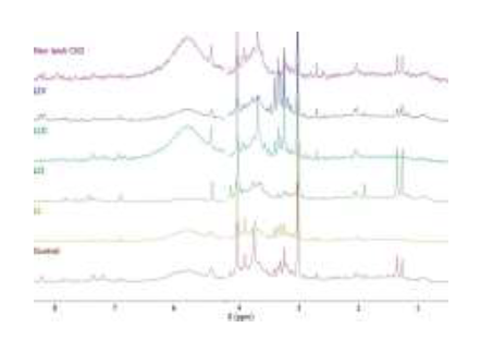
Chronic kidney disease (CKD) is a major complication and the leading cause of mortality in canine leishmaniasis (CanL). The kidneys are essential for numerous metabolic processes, and specific metabolites may serve as predictive biomarkers of kidney function. Nuclear Magnetic Resonance (NMR) spectroscopy is a prominent analytical tool in metabolomics, capable of identifying metabolites in urine. This study aim to identify distinct patterns in the NMR spectra of urine samples from dogs with CKD in CanL, reflecting the underlying metabolic profiles Fifty-five dogs were divided into three groups: 14 healthy control dogs (CG), 33 dogs with CKD secondary to leishmaniasis, and 8 dogs with CKD unrelated to leishmaniasis. CanL dogs were classified according to the International Renal Interest Society (IRIS) staging system: stage 1 (15 dogs), stage 2 (10 dogs), stage 3 (6 dogs), and stage 4 (2 dogs); and by LeishVet guidelines: stage I (5 dogs), stage II (4 dogs), stage III (14 dogs), and stage IV (10 dogs). Among dogs with CKD alone, one dog was in IRIS stage 1, two in stage 2, one in stage 3, and four in stage 4. Low-field proton nuclear magnetic resonance (1H NMR) spectroscopy and multivariate analysis were used to classify urine samples. Statistical analysis was conducted on hematology, urine and plasma samples from studied dogs. Using 1H NMR spectroscopy to classify urine samples from dogs with CKD, both with and without leishmaniasis, revealed distinct spectral patterns between the different groups. In conclusion, low-field 1H-NMR spectroscopy demonstrated that CKD presents a distinct metabolic profile compared to kidney damage secondary to leishmaniasis.